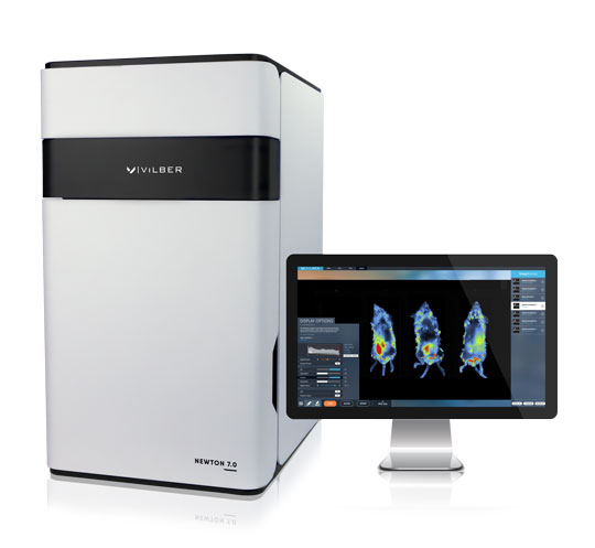
Imavita is proud to announce the recent acquisition of a new Bioluminescence imaging (BLI 2D/3D) / Fluorescence imaging (FLI 2D) imaging apparatus to help in development of preclinical models.
Since December 2022, Imavita is working on this innovative imaging system (Newton 7.0 / Vilber) which will permit to generate non-invasive, bioluminescence imaging (BLI) and fluorescence imaging (FLI) quantitative data for pre-clinical models in place at Imavita in Applications of interest, and principally in the area of drug disposition (PK and PK/PD), oncology, osteo-articular disease and dermatology.
Bioluminescence imaging / BLI is available in 2D, but also in 3D (BLI tomography). Fluorescence imaging (FLI) is available in 2D.
This new apparatus will permit in vivo and ex vivo imaging for:
- Monitoring the development of tumors and infections
- Tracking cell migration
- Targeting Biodistribution of drugs, molecules and nanoparticles
- Visualization of vasculature and microcirculation
The benefits of this in vivo imaging modality are:
- Easily and efficiently obtain calibrated quantitative data in preclinical models
- Deepen understanding of disease mechanisms, disease progression and therapeutic responses
- Manage costs and capture time course data by avoiding sacrifice of animal models
For more informations, do not hesitate to contact us.
