
| Evaluation | Parameter | Group sham (no C.acnes injection / PBS) | Test group (C.acnes injection) |
|---|---|---|---|
| In vivo | Bodyweight on 5 days | NE (No Effect) | NE or slight decrease |
| Ear thickness (calipering) | NE | Ear thickness increase <24h Stable until D5 |
|
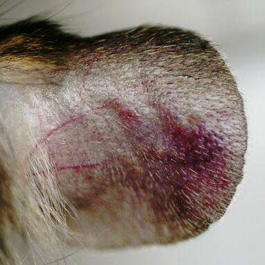
| Ear skin erythema (visual scoring or SkinColor®) | NE | Max. at +6h/+24h decreasing until Day 5 |
|
| Ear skin scaling (visual scoring) | NE | Absent or slight increase (dry skin appearance) |
|
| Ears digital pictures |  | 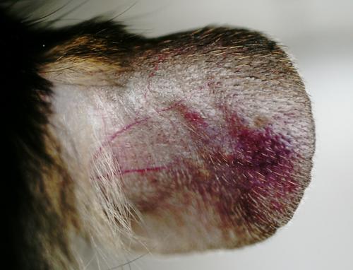 |
|
| OCT / Optical Coherence Tomography Day 5 | 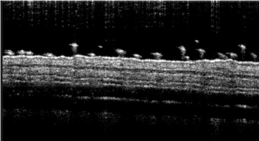 | 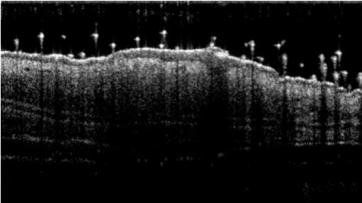 |
|
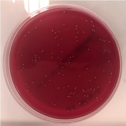
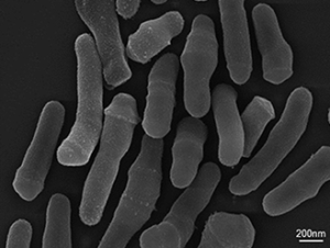
| Ex vivo | Bacteriology | Absence of C.acnes | Presence of C.acnes Numbering in agar plates |
| Genes expression qPCR (Ear / Day 5) | Normalized expression vs. sham: IL1A++ IL1B+++ MMP9+ TNFalpha++ TLR2++ |
||
| Cytokines assay Multiplexed ELISA (Ear / Day 5) | Comparison vs. sham: IL18+++ IL1B+++ TNFalpha+++ IL6+++ CXCL1+++ |
||
| Histology HES (Overview / Day 5) | 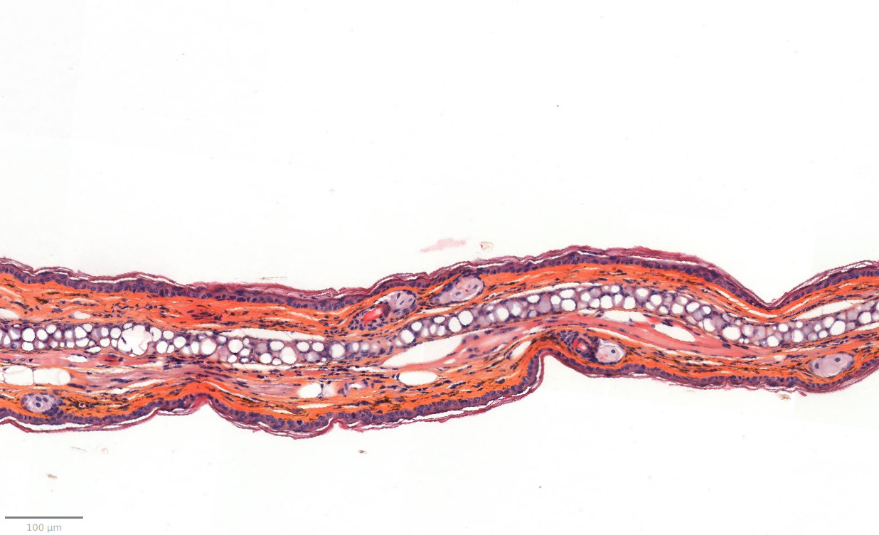 | 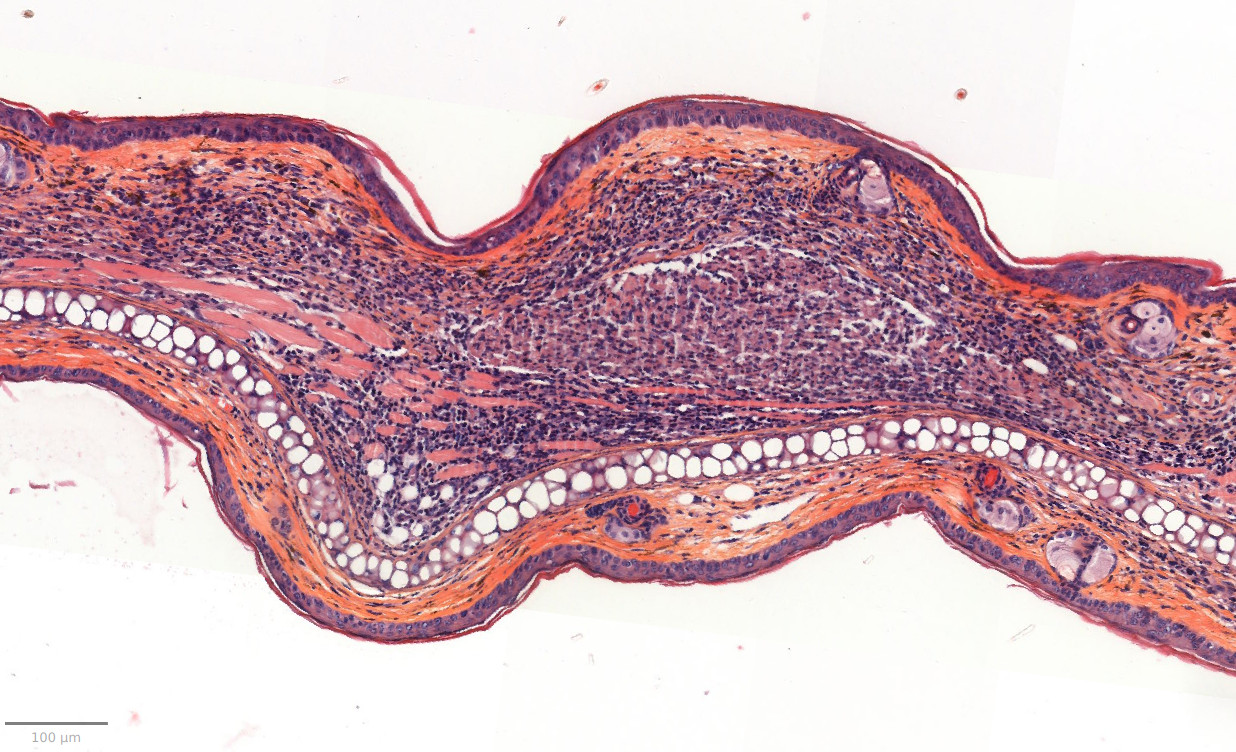 |
|
| Histomophometrics HES / Day 5 | Comparison vs. sham: Dermis inflammation (cellularity) +80-100% Dermis inflammation (tickness) +250-300% Epidermis thickness +90-100% Other: Parakeratosis / Rare presence of neutrophilic abscess (epidermis / dermis) |
||
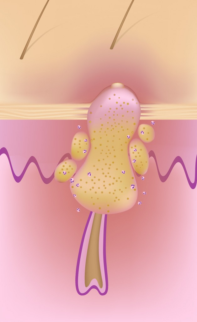
01 - Bacteria strain:
02 - Inoculum / Induction:
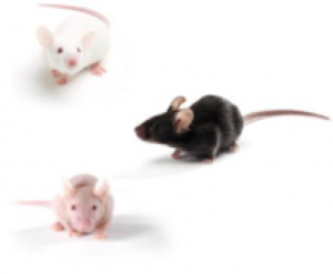
03 - Animal strain:
04 - Application of dermatological pharmaceutical products:
05 - Evaluation of in vivo clinical macroscopic signs:
06 - In vivo imaging:
07 - Ex vivo evaluations: