

In vivo monitoring of chronic skin inflammation using OCT in preclinical dermatology models
OCT in preclinical dermatology models uses near-infrared light to measure skin layers in vivo, without biopsy. In an oxazolone-induced atopic dermatitis model in hairless SKH-1 mice, epidermal thickness measured by OCT corresponded strongly to histology, with about 5 minutes per scan instead of several weeks for histological analysis.
Chronic inflammatory skin diseases, such as atopic dermatitis, psoriasis, acne, or pemphigus vulgaris, significantly impact patients’ quality of life and overall health. To better understand these conditions and evaluate potential treatments, researchers rely on preclinical dermatology models, mostly in mice. Traditionally, they require post-mortem sampling. Imavita proposes an innovative shift: applying Optical Coherence Tomography (OCT), a non-invasive imaging technique, to perform in vivo skin lesion assessment. Discover our results, presented at AFSTAL, La Rochelle, France.
The study at a glance:
| Item | Detail |
|---|---|
| Model | Oxazolone-induced atopic dermatitis in hairless SKH-1 mice |
| Protocol | Sensitization on day 1 (control group: ethanol); 11 challenges with treatments and clinical evaluations from day 8 to day 29; final measurements, clinical scoring and tissue collection on day 30 |
| OCT readout | Epidermal thickness measured in vivo at multiple time points |
| Reference method | Histology of skin sections |
| Result | Strong correspondence between OCT and histology; epidermal thickening visualized in inflamed areas |
| Speed | About 5 minutes per scan, against several weeks for histological analysis |
| Origin | Communication presented at the 45th AFSTAL Colloquium (La Rochelle, France, 2019) |
OCT reports tissue structure and thickness. Immune cell infiltration, cytokine levels and lipid barrier status still require histology or biomarker assays, and these results come from one mouse model.
The clinical context: High prevalence of inflammatory skin diseases
Inflammatory skin diseases are highly prevalent and burdensome, both clinically and socially.
Atopic dermatitis (AD) affects 10–30% of children, depending on the population studied. This makes it one of the most common pediatric dermatological conditions worldwide. Its impact extends into adulthood, with nearly 10% of individuals aged 20–30 affected (Vidal), highlighting its chronic and relapsing nature. Clinically, AD is characterized by erythematous, excoriated plaques associated with intense pruritus, leading to disrupted sleep, psychological distress, and impaired quality of life.
Another common inflammatory skin condition is psoriasis. Although less prevalent than atopic dermatitis, it affects 1–3% of the global population, including both children and adults, and remains a considerable public health concern. In addition to its cutaneous manifestations, psoriasis is associated with serious systemic comorbidities. Approximately 20% of patients develop severe forms, often accompanied by joint involvement (psoriatic arthritis), which significantly worsens patient outcomes (Inserm). Chronic inflammation and social stigma linked to the disease, further contribute to mental health deterioration in affected individuals.
Given the burden and complexity of these diseases, there is a pressing need for innovative preclinical models to advance our understanding and accelerate the development of effective therapies.
Advantages of Optical Coherence Tomography (OCT) in Preclinical Dermatology Models
OCT offers multiple advantages over conventional techniques:
- High-resolution anatomical imaging of skin layers
- Non-contact and painless for the animal
- No tissue destruction or biopsy required
- Real-time data acquisition (~5 min per scan)
- Repeated measurements possible on the same subject
- Reduced number of animals per study
- Ethically aligned with the 3Rs principle (Reduction, Refinement)
These features make OCT ideally suited for longitudinal, in vivo evaluation of inflammatory skin lesions.
Optical Coherence Tomography (OCT): A powerful imaging technology
Optical Coherence Tomography is a relatively recent, non-invasive medical imaging technique. Comparable to ultrasound, it uses near-infrared light instead of sound waves. OCT enables high-resolution visualization of the superficial layers of the skin, reaching several millimeters in depth.
How it works?
An electromagnetic wave (in the near-infrared range) is partially reflected by biological tissues. These reflections are compared with a reference beam, and the data is processed to reconstruct highly detailed cross-sectional images, with a resolution of a few microns.
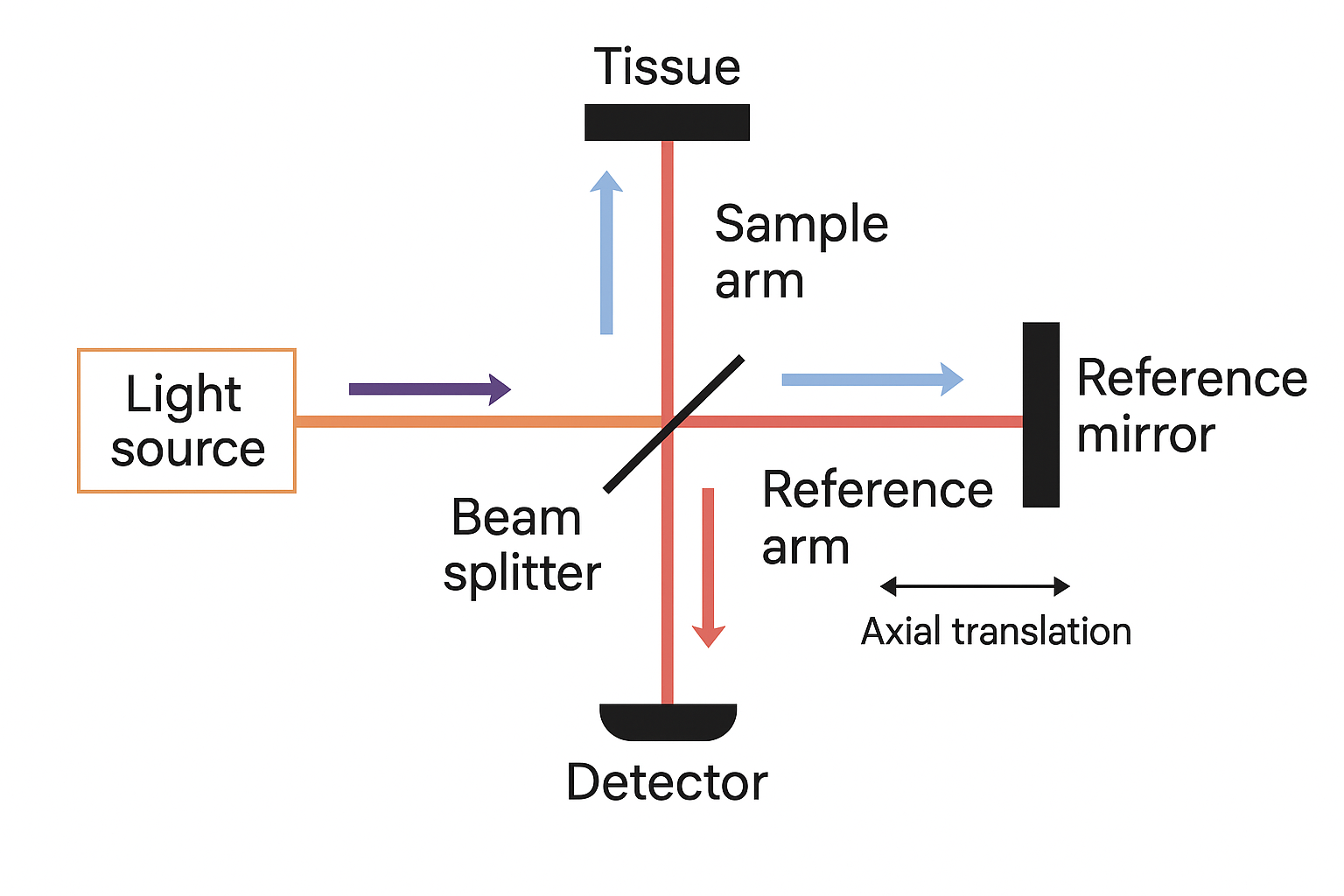
Physical principle of OCT: the Michelson interferometer
OCT is based on the principle of the Michelson interferometer, an optical setup that measures interference patterns between electromagnetic waves.
Light from the source is split into two beams:
- One beam travels through the reference arm, is reflected by a mirror, and passes back through the beam splitter.
- The other beam travels into the sample arm, where it is reflected by tissue structures.
When the two beams are recombined, they produce interference patterns. Analyzing these patterns allows for the highly precise visualization of tissue microarchitecture, with a resolution of a few microns.
Limitations of traditional approach to validating skin inflammation models
Preclinical skin models are typically assessed using post-mortem histology, which requires sacrificing animals and delays results by several weeks. Validation is based on parameters such as:
- Epidermal and dermal thickness
- Hyperkeratosis, Parakeratosis
- Lipid barrier status
- Immune cell infiltration
- Cytokine levels
While precise, these approaches lack the ability to track changes dynamically over time in the same subject.
Imavita’s experimental: A non-invasive shift
Imavita implemented OCT in a validated oxazolone-induced atopic dermatitis model in hairless SKH-1 mice.
Protocol and study timeline:
- Day 1: Sensitization with oxazolone (control group: ethanol)
- Days 8–29: 11 challenges + treatments + clinical evaluations
- Day 30: Final measurements, clinical scoring, and tissue collection
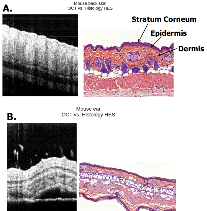
Epidermal thickness was measured in vivo at multiple time points using OCT. These measurements were then compared to traditional histological analyses (microscopic examination of skin sections).
Comparison of tomography and histology results: Notable results on the benefits of tomography compared with histology
- Strong correspondence between OCT and histological measurements
- OCT enables rapid follow-up (5 minutes per scan), compared with several weeks for histological analysis
- Effective visualization of epidermal thickening in inflamed areas
Why is OCT a turning point for animal models?
This imaging technique offers a more ethical approach, aligned with the 3Rs principles adopted by Imavita, while also being more efficient and scientifically robust.
- Fewer animals required
- Dynamic follow-up over time on the same subject
- No pain or heavy anesthesia
- Immediately usable results
Perspectives: expanding the use of Optical Coherence Tomography to other research areas
Following its success with atopic dermatitis, other models are currently being validated at IMAVITA:
- Psoriasis model in mice
- Pemphigus vulgaris in mouse
- Medical device validation (micro-needles)
- Ophtalmology / ocular disease
- Wound healing models
- Potential expansion to oncology
Optical Coherence Tomography has the potential to transform preclinical dermatological research. Non-invasive, fast, and repeatable, it offers a powerful alternative to conventional histological methods. This marks a step toward more modern, 3Rs-compliant, and efficient science.
Discover our advanced preclinical imaging solutions
Frequently asked questions about OCT in preclinical dermatology models
What is OCT imaging in preclinical dermatology?
Optical coherence tomography is a non-invasive technique that uses near-infrared light, in the way ultrasound uses sound waves, to image the superficial layers of the skin with a resolution of a few microns and no biopsy.
How well do OCT measurements match histology?
In Imavita’s oxazolone-induced atopic dermatitis model in SKH-1 mice, OCT and histological measurements of epidermal thickness showed strong correspondence, and OCT showed epidermal thickening in inflamed areas.
How does OCT support the 3Rs?
OCT allows repeated scans of the same animal without biopsy or euthanasia, which supports Refinement and can reduce the number of animals per study. See the 3Rs in preclinical studies.
What does OCT not replace?
OCT reports tissue structure and thickness. Immune cell infiltration, cytokine levels and lipid barrier status still require histology or biomarker assays.
Which models can OCT be applied to?
OCT was validated in the oxazolone-induced atopic dermatitis model. Other models are being validated at Imavita: psoriasis in mice, pemphigus vulgaris in mouse, medical device validation (micro-needles), ophthalmology and ocular disease, wound healing models, with a potential extension to oncology.
Supporting scientific evidence
The relevance of OCT in skin research is also supported by the literature:
- Wu et al. (2012) demonstrated that OCT can quantify epidermal thickness and optical attenuation coefficients, distinguishing between chronological and photoaged skin in mice.
- Schuetzenberger et al. (2020) showed that OCT allows longitudinal, volumetric assessment of intradermal hydrogel implants, confirming its precision and repeatability for tracking tissue changes over several months.
These findings align with Imavita’s observations and reinforce the scientific legitimacy of OCT in preclinical dermatology models.
Contact Imavita to explore how Optical Coherence Tomography can enhance your dermatology research models.
Sources:
Communication presented at the 45th Afstal Colloquium (La Rochelle, France, 2019)
Eric Lacoste et al. IMAVITA, Canal Biotech 1, Parc Technologique du Canal, 3 rue des Satellites, 31400 TOULOUSE, FRANCE.
References:
Wu S, Li H, Zhang X, Li Z. Optical features for chronological aging and photoaging skin by optical coherence tomography. Lasers Med Sci. 2012;28(2):445-450. doi:10.1007/s10103-012-1069-4
Schuetzenberger K, Pfister M, Messner A, Garhöfer G, Hohenadl C, Pfeiffenberger U, Schmetterer L, Werkmeister RM. Cutaneous optical coherence tomography for longitudinal volumetric assessment of intradermal volumes in a mouse model. Sci Rep. 2020;10:4245. doi:10.1038/s41598-020-61276-9