A human three-layer skin explant model of Cutibacterium acnes infection: inflammation, matrix remodeling and adipose response
Key takeaways
- Imavita and DIVA Expertise infected human full-thickness skin explants (epidermis, dermis and subcutaneous fat) with Cutibacterium acnes by intradermal injection.
- Live bacteria persisted for 72 h, and the explants mounted an NF-κB and inflammasome-driven cytokine response (TNF-α, IL-1α, IL-6, IL-8).
- The model also captured extracellular matrix remodeling and changes in adipocyte metabolism, a compartment absent from most acne models.
- Results are from 3 donors and were presented as a poster at the 42nd CARD congress (Toulouse, 25–26 June 2026).
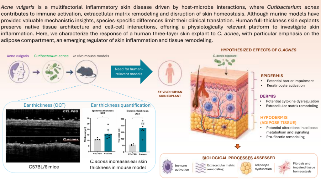
Why a human skin explant model for acne?
Acne vulgaris is a chronic inflammatory disease of the pilosebaceous unit in which C. acnes contributes to immune activation, matrix remodeling and loss of skin homeostasis. Mouse models remain useful for mechanism and efficacy, but mouse skin differs from human skin in thickness, follicle density and immune composition. Human full-thickness explants keep the native architecture and cell-cell interactions, including the subcutaneous adipose tissue now recognized as an active player in skin inflammation.
Study design / Material & Method
Explants origin
Skin was obtained from abdominal dermolipectomy of three female donors (35, 52 and 38 years; BMI 24–26.3 kg/m²) and prepared as three-layer explants (DIVA skin).
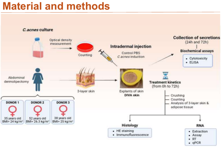
Induction
Explants received an intradermal injection of C. acnes (8.19 × 10⁷ CFU in 40 µL) or PBS.
Culture supernatants were collected at 24 h and 72 h for cytotoxicity and ELISA.
Analysis
Tissues were analyzed by:
- Bacterial counting
- Histology (H&E, immunofluorescence)
- RT-qPCR.

Results
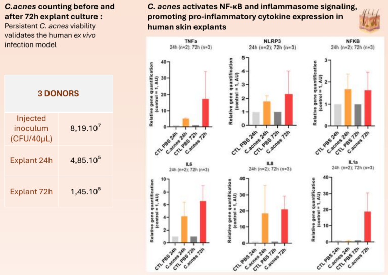
The infection is real and sustained
Viable C. acnes were recovered from explants at 24 h (4.85 × 10⁵ CFU) and 72 h (1.45 × 10⁵ CFU), confirming that the model tests a live host-microbe interaction, not a bacterial lysate.
Inflammation through NF-κB and the inflammasome
C. acnes increased expression of NFKB and NLRP3, and of TNF-α, IL-1α, IL-6 and IL-8. At 72 h, TNF-α and IL-1α reached roughly 17- to 19-fold control levels (n = 2 donors at 24 h, n = 3 at 72 h).
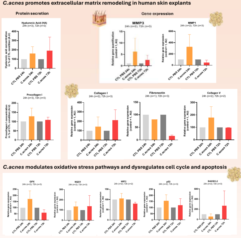
Extracellular matrix remodeling
MMP1 and MMP3 expression rose early (24 h). Hyaluronic acid secretion and collagen I expression increased, while fibronectin decreased at 72 h.
Oxidative stress and cell fate.
GPX, NQO1, p53 and the Bax/Bcl-2 ratio increased, whereas NRF2 decreased, pointing to oxidative stress and altered cell-cycle and apoptosis control.
The adipose compartment responds.
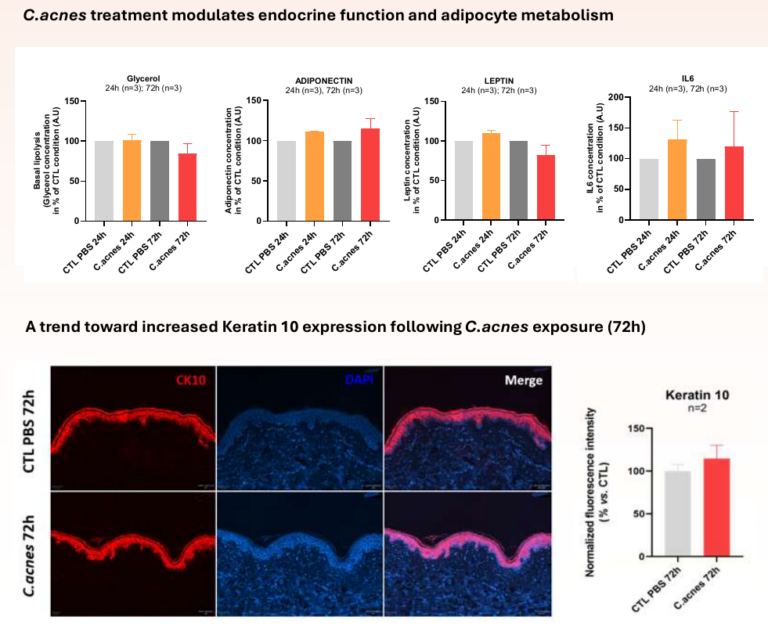
Leptin and glycerol release decreased and adiponectin rose slightly at 72 h, suggesting that C. acnes modulates adipocyte metabolism and the skin secretome. Keratin 10 immunostaining showed a trend toward higher expression (n = 2).
Features
How this fits with in vivo work
Imavita runs the ex vivo human model alongside an in vivo C. acnes ear model in C57BL/6 mice. In that model, optical coherence tomography (OCT) measures epidermal and dermal thickening non-invasively and longitudinally, in the same animals. A sponsor can screen candidates on human tissue first, then confirm efficacy in vivo with fewer animals, in line with the 3Rs.
Limitations
These are exploratory data: three donors, two time points, no formal statistical testing shown, and a donor profile limited to women with abdominal skin. Explants lack circulating immune cells and sebaceous-rich facial skin, so they model the tissue response to C. acnes, not the full clinical disease. Findings will be extended to more donors and to reference treatments.
Our posters related to use cases are available on request
Discover more on our services
FAQ zone
What is a three-layer skin explant?
A piece of human skin that keeps the epidermis, dermis and subcutaneous fat, maintained alive in culture for several days.
Which readouts are available?
Bacterial load (CFU), secreted cytokines and adipokines (ELISA), gene expression (RT-qPCR), histology and immunofluorescence, and cytotoxicity.
Can I test my compound in this model?
Yes. Topical or injected treatments, reference comparators (e.g. antibiotics, retinoids) and custom readouts can be included. Contact us to define the design.
Is there an in vivo follow-up?
Yes, the C57BL/6 ear model with OCT imaging, plus histology and cytokine analysis.